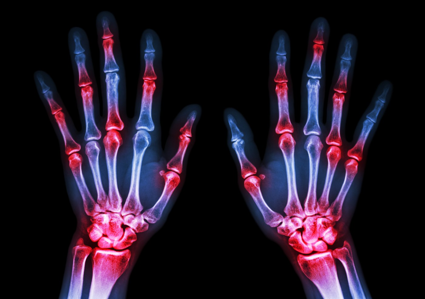
 A study recently published in the journal Arthritis showed that digital X-ray radiogrammetry can be used as an imaging tool to assess bone mineral density loss in patients with rheumatoid arthritis (RA). The study is entitled “Early metacarpal bone mineral density loss using digital x-ray radiogrammetry and 3-tesla wrist MRI in established rheumatoid arthritis: a longitudinal one-year observational study.”
RA is an autoimmune disease that causes chronic inflammation and pain in the joints and other parts of the body due to an overreaction of the body’s own immune system, which results in the attack of healthy tissues. Radiographic imaging, or X-ray, has been used in RA diagnosis and to routinely evaluate disease progression in terms of structural damage, especially in patients’ hands and feet. Newer imaging techniques, like magnetic resonance imaging (MRI), play an important role primarily in detecting early changes such as erosions, synovitis (inflammation in the lining of a joint) and bone marrow edema (BME — when excess fluids accumulate in the bone marrow causing swelling and inflammation).
RA patients usually exhibit, as one of the early radiographic changes, periarticular osteopenia, a bone condition characterized by a decrease in bone mineral density leading to bone weakening and increased risk of bone fractures. The loss of metacarpal bone (specific bones in the hand) mineral density is a known predictor of RA development.
A new automated radiogrammetric method has been previously developed to evaluate bone mineral density loss based on single radiographs from hands. The goal of this study was to assess the relationship of the digital X-ray radiogrammetry (DXR) in the evaluation of early metacarpal bone mineral density (BMD) loss, disease activity by high field strength 3-tesla wrist MRI, and radiographic changes in the hands and feet of RA patients. To this end, 10 RA patients were assessed who had hand and feet X-rays and wrist MRI at several time points during a follow-up period of one year.
Researchers found a strong correlation between early changes in BMD at 12 weeks and changes in wrist BME after 1 year of follow-up, but not with synovitis, erosions or radiographic scores. No significant changes were found in disease activity based on wrist MRI and total hand and feet X-ray scores in this patient cohort.
The research team concluded that early changes in BMD correlated with changes observed after one year in wrist BME, a predictor of future erosion and joint damage in RA, but not with the progression of radiographic damage or MRI erosion in the cohort of patients analyzed. Researchers hypothesized that this might be due to a slower manifestation of MRI/radiographic erosions in comparison with BMD changes, suggesting that early changes in BMD may actually represent a more sensitive prognostic marker of RA in the follow-up period. The research team proposes that DXR may be used as a tool for early detection of BMD and damage progression, enabling clinicians to use a low-cost, readily available technique to follow-up RA patients in comparison with MRI.


