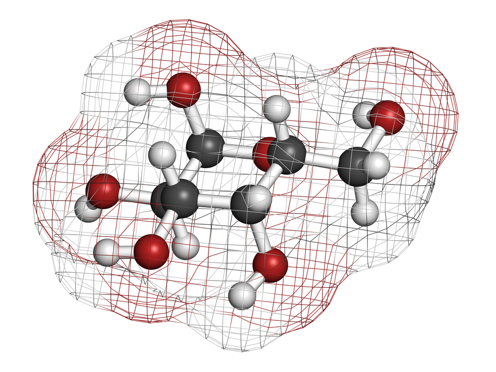
Researchers agree that interactions between cell membrane proteins and sugars hold the key to understanding the molecular basis of many diseases. Sugar molecules, which are attached to cell membranes like an antennae, are responsible for interactions with foreign molecules, transfer of signals across membranes, and regulation of physiological processes. Most diseases are caused due to communication errors between these molecules as a result of mutations in these proteins or the structure of sugar molecules being altered. As a result, studying the interaction of these sugar molecules in vitro would give a clear insight to the molecular basis of disease and help in formulating appropriate therapeutic strategies for each.
Since the cell membrane is made of phospholipids with a hydrophilic head and a hydrophobic tail, the easiest way to obtain a laboratory model of the cell membrane would be to make the phospholipid forming units (molecules) self-assemble themselves in water. However, the product of this process is very short lived and dissociates easily.
To study these interactions extensively, in 2010, a group of researchers lead by Virgil Percec, a professor of chemistry in the University of Pennsylvania School of Arts and Sciences, along with postdoctoral chemistry researchers Shaodong Zhang and Ralph-Oliver Moussodia in collaboration with Temple University’s Michael Klein, as well as Sabine Vértesy and Sabine André and Hans-Joachim Gabius of Ludwig-Maximillians University in Munich devised an artificial form of membrane with the help of amphiphilic Janus dendrimers, which have water-loving and water-hating branches, instead of heads and tails. Unlike before, when the positioning of sugar molecules and their modification used to be almost impossible, these dendrimers made their task less tedious. These dendrimers could be bonded to glycans (forming glycodendrimers), which could in turn be diluted in a series of organic solvents and then injected in water to produce the simplest cell membranes with various combinations of glycans (sugars).
“As our molecules self-assemble, the vesicles formed have a precise number and spatial arrangement of the sugars, something never possible before,” Percec said. Their work was published in the 2013 edition of the Journal of the American Chemical Society.
Recently, according to a press release by The University of Pennsylvania, these scientists have tested the molecular interactions of the Gal-8 (galectin-8) protein — an important cell-signaling protein which when mutated, can cause rheumatoid arthritis in many patients. A single change in the building block of this protein impaired its ability to interact with the artificial membrane which could be the molecular basis of rheumatoid arthritis.
“By testing this model with a sugar binding protein of human origin, we show that single mutation of an amino acid from a giant protein structure can induce a dramatic change in its interactions with the cell,” Percec said. “This demonstrates just how efficient and sensitive a model this is for biological membranes.”
The study was published in The Proceedings of the National Academy of Sciences.
In the future, these artificial dendrimers and their interaction with different molecules apart from Gal-8 would shed light on the molecular basis for many diseases which until now were poorly understood.


